
What is Live and Dry Blood Analysis?
Live Blood Analysis is used in the field of complementary medicine, often in conjunction with nutrition, naturopathy, homeopathy, acupuncture or herbalism.
A drop of the client’s blood is examined under a microscope and the information gained is used to check for signs of many issues relating to health. We are able to use it to bring a new dimension to our ability to understand what is happening at a cellular level within our clients.
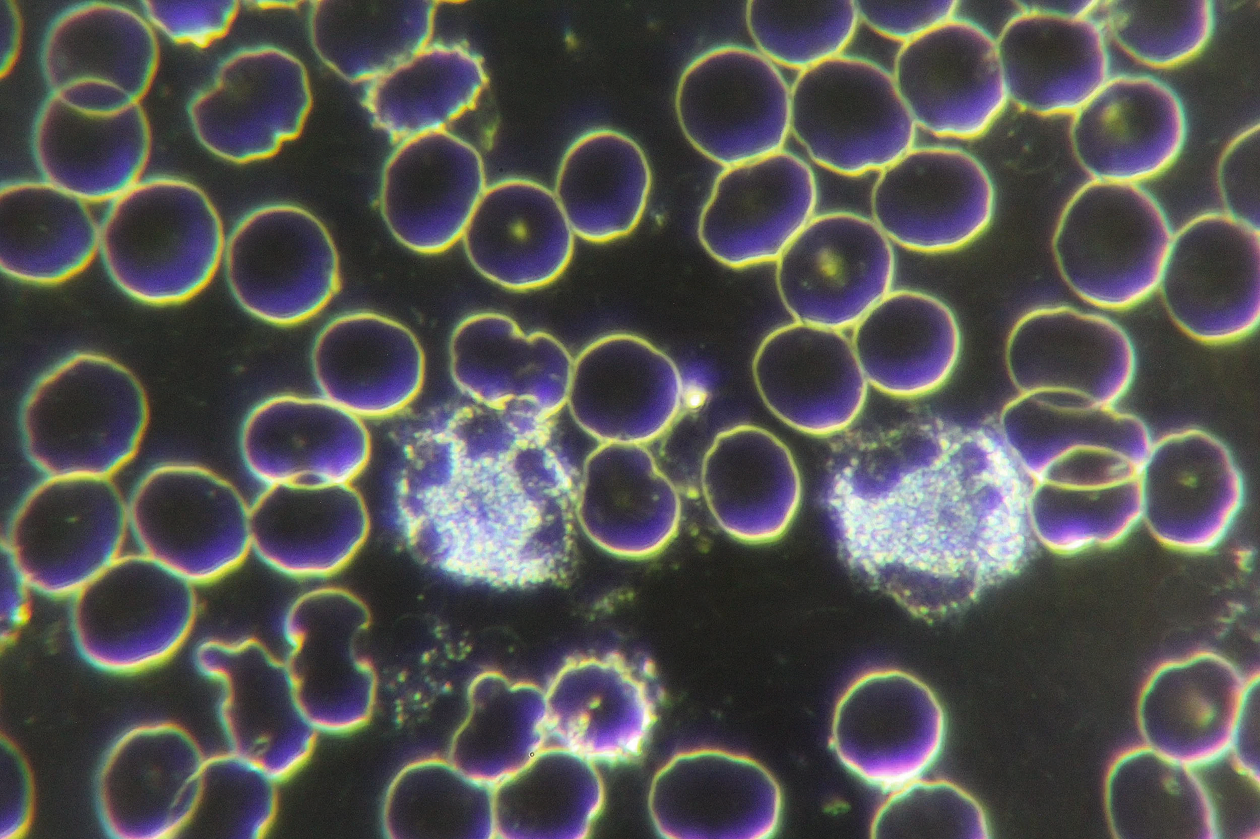
Live Blood Analysis
Why is it called ‘live’ blood analysis?
An LBA blood sample is not stained or treated with chemicals - it is mounted onto the microscope as soon as possible after being extracted to be able to view it in its ‘live’ state.
This enables us to see how the blood moves, how the blood cells react to each other and how they respond to the environment they float in – the plasma.
By magnifying the blood 1000 times, we can see the health of each individual cell and are able to assess how many cells are normal and functional and how many cells are not. Abnormal cells are obvious and their presence will indicate a variety of issues.
What can a Live Blood Analysis show?
One drop of blood contains 5 million cells. The proportions and amounts of the different cells distributed throughout that one drop is representative of what is contained in all of a person’s blood.
From this one drop of blood we can:
Determine if there are specific deficiencies of nutrients such as B12, folate, zinc, Vitamin C, or EFAs.
Check if the cell membranes are damaged or whether they are healthy.
Assess immune system cell activation and whether there are enough/too many white blood cells - this can show the presence of inflammation, autoimmune issues, infection, allergies or parasites.
Check for elements in the plasma that can indicate liver issues or circulatory challenges, such as thrombocytes, uric acid crystals or fat crystals.
Assess the integrity of the digestive tract and screen for problems such as ‘leaky gut’.
Assessment of the charge of the red blood cell membranes – the zeta potential – which is indicative of the pH of the fluids/tissues of the body as a whole
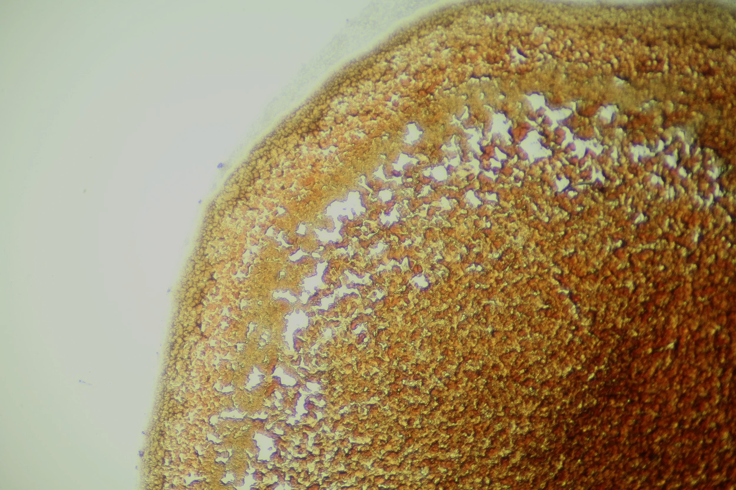
The Dry Blood Analysis procedure involves puncturing a client’s fingertip and allowing a bead of blood to form. This is left on the finger for 30 seconds to allow the clotting cascade to develop. A microscope slide is then pressed onto the drop of blood eight times. The slide is left to dry, which takes about 15 minutes, and in that time the eight blood layers will go through an extraordinary transformation.
As blood is a biological fluid - it behaves like a non-Newtonian fluid. This means that as the water evaporates from layers of blood it generates currents within the fluid. This moves the contents of the layer around and creates the distinctive patterns.
How the contents move and where they end up within the layer is determined by the electro-biochemical properties of the red blood cells, the quantity of the various clotting factors in the plasma, toxins that have been circulating within the blood, by-products of pathogens, by-products of oxidation, free radical damage and degenerative disease processes. This is what generates the distinctive patterns within the dry blood layers that are indicative of various issues going on within the body.
Dry Blood Analysis
What can Dry Blood Analysis show?
Detect the presence of inflammation
Assess the progression of inflammatory processes - chronic or acute
Determine the location of the inflammatory process – organ, tissue or system specific
Monitor the progression or reduction of the inflammatory process
Detect heavy metal toxicity
Reveal the presence of parasites
Show mineral and vitamin imbalances
Show antibiotic damage
Detect bowel inflammation
Assess the lymphatic burden
Assess protein metabolism